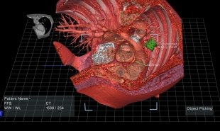
Doctors currently rely on flat images from CT and MRI scans for pre-op information about patient’s organs. Now, however, health tech startup EchoPixel is planning to use the information garnered from current medical imaging technology to produce 3D virtual reality organs, which doctors can explore and inspect before beginning surgery.
EchoPixel uses the images which are already being gathered during medical imaging processes to create 3D-rendered body parts. These floating masses can then be examined via a VR platform called zSpace. Doctors can rotate and dissect the images of organs, including the brain and the heart, using a stylus. They can even examine a colon via a simulated fly-through.

EchoPixel hope their technology will help doctors gain an enhanced understanding of the intricacies of each organ, and enable them to go into surgery well-rehearsed. It can also be used by medical students as a supplementary learning tool. Could this combined technology be used in other industries too — such as mechanics or construction?
Website: www.echopixeltech.com
Contact: www.echopixeltech.com/contact-us


